
How is femoral anteversion diagnosed?
The diagnosis of femoral anteversion is made by a physical examination by your child’s physician. During the examination, the physician obtains a complete prenatal and birth history of the child and asks if other family members are known to have femoral anteversion.
Other diagnostic procedures may include:
Other diagnostic procedures may include:
- x-ray – a diagnostic test which uses invisible electromagnetic energy beams to produce images of internal tissues, bones, and organs onto film.
- computed tomography (Also called CT or CAT scan.) – diagnostic imaging procedure that uses a combination of x-rays and computer technology to produce cross-sectional images (often called slices), both horizontally and vertically, of the body. A CT scan shows detailed images of any part of the body, including the bones, muscles, fat, and organs. CT scans are more detailed than general x-rays.
- magnetic resonance imaging (MRI) – a diagnostic procedure that uses a combination of large magnets, radiofrequencies, and a computer to produce detailed images of organs and structures within the body.
In addition, some physicians place ink or chalk on the bottom of the child’s feet and have them walk on paper to evaluate the amount of intoeing the child has.
Long-term outlook for a child with femoral anteversion
Femoral anteversion has a very good prognosis. Many cases correct themselves as the child grows. On rare occasions, femoral anteversion can be severe and surgery may be required to straighten the thigh bone.
It is important to know that femoral anteversion typically does not lead to arthritis or any other future health problems
It is important to know that femoral anteversion typically does not lead to arthritis or any other future health problems
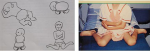
- Exacerbated by sitting in ‘W’ position
- More common in girls
- Many correct spontaneously by age 10 years
5-10% persist , but rarely require surgical correction